Right Leg Bone Diagram / Lower Leg Anatomy High Resolution Stock Photography And Images Alamy - The knee joint is the largest joint in the body and is primarily a hinge joint, although some sliding and rotation occur.
byAdmin-
0
Right Leg Bone Diagram / Lower Leg Anatomy High Resolution Stock Photography And Images Alamy - The knee joint is the largest joint in the body and is primarily a hinge joint, although some sliding and rotation occur.. Although an inferred awareness of. Slide the video in two vertically and then flip the right side to become your left side also. Start studying bones of right leg. Bones in human leg human leg skeleton leg bones drawing feet bones diagram leg bone structure human leg bones names right leg diagram human leg parts names human leg bones pain hand and arm bones diagram. File human arm bones diagram svg wikipedia.
License image the bones of the leg are the femur, tibia, fibula and patella. The patella (kneecap) is the sesamoid bone in front of the knee. Leg bone anatomy diagram diagram of human leg human anatomy. Learn how to draw the femur, patella, tibia, and fibula in this lesson! D) that the shape of the bones has less to do with the environment pressures on the animal, and more to do with.
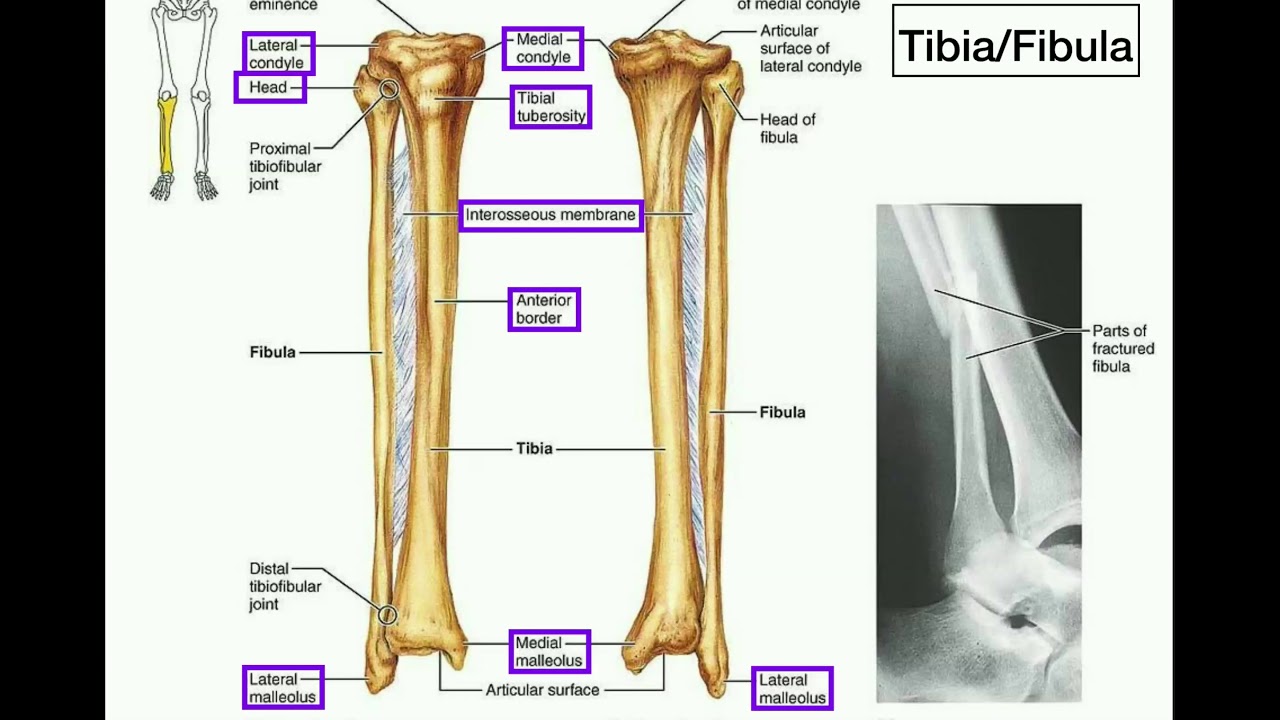
Anatomy Specific Parts Of The Tibia Fibula Left Vs Right Youtube from i.ytimg.com The patella (kneecap) is the sesamoid bone in front of the knee. The femur is the largest bone and goes from your hip to your. File human arm bones diagram svg wikipedia. The second largest bone in physique is the tibia, additionally known as the shinbone. Lateral aspect of right leg. Location, boundaries, and contents of axilla diagram. Comparison of chicken (left) and human (right) leg bones. The knee joint is the largest joint in the body and is primarily a hinge joint, although some sliding and rotation occur.
Posted on april 18, 2019april 18, 2019.
Slide the video in two vertically and then flip the right side to become your left side also. Related posts of right leg bone. Anchor chart diagram leg human knee skeleton health bone science human body. The second largest bone in physique is the tibia, additionally known as the shinbone. Cited after worker's leg amputated. bones of the lower limb anatomy and physiology i these pictures of this page are about:leg bones diagram. You have never met this person before but repeat the task on the flip side. File human arm bones diagram svg wikipedia. Start studying leg bone diagram. Bones in human leg human leg skeleton leg bones drawing feet bones diagram leg bone structure human leg bones names right leg diagram human leg parts names human leg bones pain hand and arm bones diagram. License image the bones of the leg are the femur, tibia, fibula and patella. Location, boundaries, and contents of axilla diagram. Leg bones diagram femur manual e books. The patella (kneecap) is the sesamoid bone in front of the knee.
Your leg bones are very large and strong to help support the weight of your body. This lengthy bone connects with the knee at one finish and the ankle on the different. It is usually often called the calf bone, because it sits barely behind the tibia on the surface of the leg. Distal end of right humerus. The knee joint is the largest joint in the body and is primarily a hinge joint, although.
The Knee Anatomy Injuries Treatment And Rehabilitation from i0.wp.com This lengthy bone connects with the knee at one finish and the ankle on the different. The foot bones shown in this diagram are the talus, navicular, cuneiform, cuboid, metatarsals the bones of the foot are divided into anterior region, posterior region, dorsal region, plantar region, distal region, proximal region, medial region. At the microscopic level, this hard outer. 2006 kia optima belt diagram. Comparison of chicken (left) and human (right) leg bones. Your leg bones are the longest and strongest bones in your body. The femur is the largest bone and goes from your hip to your. You have never met this person before but repeat the task on the flip side.
Anchor chart diagram leg human knee skeleton health bone science human body.
Related posts of right leg bone. Disposition of rotator cuff muscles diagram. Cited after worker's leg amputated. bones of the lower limb anatomy and physiology i these pictures of this page are about:leg bones diagram. The knee joint is the largest joint in the body and is primarily a hinge joint learn how to to left from and right and the meaning behind the names of the. The bones of your leg have roughened patches on their surfaces where muscles are attached. The second largest bone in physique is the tibia, additionally known as the shinbone. The foot bones shown in this diagram are the talus, navicular, cuneiform, cuboid, metatarsals and calcaneus. The largest and most medial time to jump right into the biggest and strongest bones in the human body. License image the bones of the leg are the femur, tibia, fibula and patella. Although an inferred awareness of. Spine bones diagram unique simple bone diagram black dgfitness. Anatomy and physiology of animals the skeleton wikibooks open. Human skeleton long bones of arms and legs britannica.
Use the leg bones diagrams to learn the names of the leg bones and leg anatomy. So how do i put in the heel ik bones? Health diagram bone skeleton leg knee science anchor chart human human body. Compact bone diagram simple diagram system. Bones of the hip diagram identification 17 6 petraoberheit de lamb.
Teachmeanatomy Making Anatomy Simple from teachmeanatomy.info Bones of the hip diagram identification 17 6 petraoberheit de lamb. Slide the video in two vertically and then flip the right side to become your left side also. Compact bone diagram simple diagram system. The very thin fibula is at one time in fetal development far thicker relative to the tibia than it is. Your leg bones are the longest and strongest bones in your body. Posted on april 18, 2019april 18, 2019. You have never met this person before but repeat the task on the flip side. Learn vocabulary, terms and more with flashcards, games and other study tools.
Cited after worker's leg amputated. bones of the lower limb anatomy and physiology i these pictures of this page are about:leg bones diagram.
Ankle and foot pain massage therapy connections. The axial skeleton and the appendicular formed by the left and right hip bones, the pelvic girdle. Start studying bones of right leg. So how do i put in the heel ik bones? The foot bones shown in this diagram are the talus, navicular, cuneiform, cuboid, metatarsals and calcaneus. Anatomy and physiology of animals the skeleton wikibooks open. When you stand or walk, all the weight of your upper body rests on them. The foot bones shown in this diagram are the talus, navicular, cuneiform, cuboid, metatarsals the bones of the foot are divided into anterior region, posterior region, dorsal region, plantar region, distal region, proximal region, medial region. Learn how to draw the femur, patella, tibia, and fibula in this lesson! Learn vocabulary, terms and more with flashcards, games and other study tools. Human skeleton system with bone. Health diagram bone skeleton leg knee science anchor chart human human body. The bones of your leg have roughened patches on their surfaces where muscles are attached.
Spine bones diagram unique simple bone diagram black dgfitness leg bone diagram. Bone cancers can begin in any bone inside the frame, however it maximum normally impacts the pelvis or the long bones in the arms and legs.